
Файл:HIV-budding-Color.jpg

Памер прагляду: 800 × 531 піксэлаў. Іншыя разрозьненьні: 320 × 213 піксэлаў | 640 × 425 піксэлаў | 1024 × 680 піксэлаў | 1280 × 850 піксэлаў | 2967 × 1971 піксэлаў.
Арыгінальны файл (2967 × 1971 піксэль, памер файла: 3,92 Мб, тып MIME: image/jpeg)
|
|
Гэты файл паходзіць зь Вікісховішча. Зьвесткі пра гэты файл зь яго старонкі апісаньня прыведзеныя ніжэй. Вікісховішча — сховішча вольных мэдыяфайлаў. Вы можаце дапамагчы. |
Апісаньне
| Апісаньне |
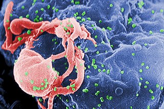
English: Scanning electron micrograph of HIV-1 budding (in green) from cultured lymphocyte. This image has been colored to highlight important features; see PHIL 1197 for original black and white view of this image.
Multiple round bumps on cell surface represent sites of assembly and budding of virions.
Español: Microfotografía con MEB de VIH-1 en liberación (en verde) en un cultivo de linfocitos. Esta imagen ha sido coloreada para resaltar las características importantes; para la imagen original en blanco y negro véase PHIL 1197. Las múltiples protuberancias redondeadas sobre la superficie celular representa los sitios de ensamblado y gemación de viriones.
Français : Virus HIV fixé sur un lymphocyte vu en microscopie électronique (fausses couleurs, le VIH est en vert).
Bahasa Indonesia: HIV yang baru memperbanyak diri tampak bermunculan sebagai bulatan-bulatan kecil (diwarnai hijau) pada permukaan limfosit setelah menyerang sel tersebut; dilihat dengan mikroskop elektron.
Русский: Фотография, полученная с помощью сканирующего электронного микроскопа. Вирусы ВИЧ (зелёные) отпочковываются от заражённого лимфоцита. Фотография была раскрашена с целью подчеркнуть важные детали; см. исходную чёрно-белую версию ниже.
Многочисленные круглые выпуклости на поверхности клетки являются местами сборки и отпочковывания вирионов.
Български: Вирусът ХИВ (в зелено) разспространяващ се от вече заразен лимфоцит.
Polski: Fotografia wykonana skaningowym mikroskopem elektronowym - przedstawia wirusy (kolor zielony) wydostających się z limfocytu. |
||
| Дата | |||
| Крыніца |
|
||
| Аўтар |
|
||
| Дазвол (Паўторнае выкарыстаньне гэтага файлу) |
PD-USGov-HHS-CDC English: None - This image is in the public domain and thus free of any copyright restrictions. As a matter of courtesy we request that the content provider be credited and notified in any public or private usage of this image. |
||
| Іншыя вэрсіі |
|


fuk12
Ліцэнзія
This image is a work of the Centers for Disease Control and Prevention, part of the United States Department of Health and Human Services, taken or made as part of an employee's official duties. As a work of the U.S. federal government, the image is in the public domain.
|
Гісторыя файла
Націсьніце на дату/час, каб паглядзець, як тады выглядаў файл.
| Дата і час | Мініятура | Памеры | Удзельнік | Камэнтар | |
|---|---|---|---|---|---|
| цяперашняя | 03:16, 20 красавіка 2008 | | 2967 × 1971 (3,92 Мб) | Optigan13 | {{Information |Description={{en|Scanning electron micrograph of HIV-1 budding from cultured lymphocyte. See PHIL 1197 for a black and white view of this image. Multiple round bumps on cell surface represent sites of assembly and budding of virions.}} |Sou |
Выкарыстаньне файла
Наступная старонка выкарыстоўвае гэты файл:
Глябальнае выкарыстаньне файла
Гэты файл выкарыстоўваецца ў наступных вікі:
- Выкарыстаньне ў ar.wikipedia.org
- Выкарыстаньне ў arz.wikipedia.org
- Выкарыстаньне ў ast.wikipedia.org
- Выкарыстаньне ў as.wikipedia.org
- Выкарыстаньне ў azb.wikipedia.org
- Выкарыстаньне ў az.wikipedia.org
- Выкарыстаньне ў bg.wikipedia.org
- Выкарыстаньне ў bn.wikipedia.org
- Выкарыстаньне ў ca.wikipedia.org
- Выкарыстаньне ў ca.wikinews.org
- Выкарыстаньне ў ckb.wikipedia.org
- Выкарыстаньне ў cs.wikipedia.org
- Wikipedie:Studenti píší Wikipedii/Pokroky v imunologii I (2013/2014)
- Wikipedie:Studenti píší Wikipedii/Pokroky v imunologii I (2014/2015)
- Wikipedie:Nástěnka/Univerzita Karlova/Pokroky v imunologii (2013-2014)
- Wikipedie:Nástěnka/Univerzita Karlova/Molekulární imunologie (2014-2015)
- Wikipedie:Nástěnka/Univerzita Karlova/Pokroky v imunologii (2014-2015)
- Выкарыстаньне ў cy.wikipedia.org
- Выкарыстаньне ў de.wikipedia.org
- Выкарыстаньне ў diq.wikipedia.org
- Выкарыстаньне ў en.wikipedia.org
- Выкарыстаньне ў en.wikibooks.org
Паказаць глябальнае выкарыстаньне гэтага файла.